

Solitary mastocytoma
Solitary cutaneous mastocytoma; single mast-cell lesion; pediatric solitary mastocytosis
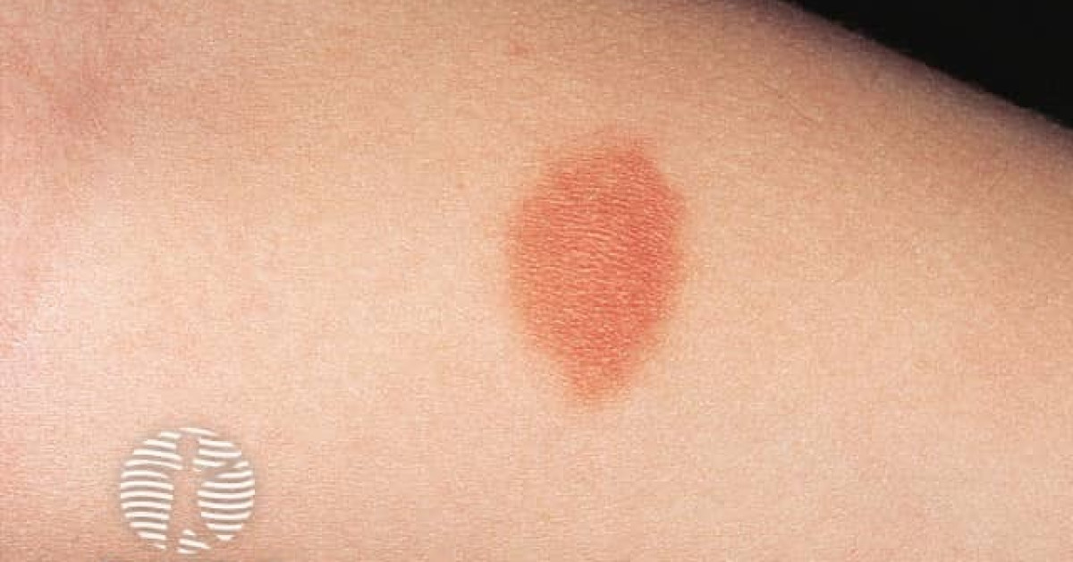
Solitary mastocytoma is one of three principal patterns of paediatric cutaneous mastocytosis — alongside maculopapular cutaneous mastocytosis (urticaria pigmentosa) and diffuse cutaneous mastocytosis. It presents in infancy / early childhood as a single tan, pink or yellow-brown plaque, often on the trunk or extremity, that develops urtication and erythema on stroking (positive Darier sign — pathognomonic). Histology shows a dense dermal infiltrate of mast cells; KIT D816V mutation is present in many but at lower prevalence than adult mastocytosis. The vast majority spontaneously regress by adolescence. Management is symptomatic — H1/H2 antihistamines, sodium cromoglycate, avoidance of mast-cell degranulators (NSAIDs, opioids, alcohol, vigorous rubbing, water temperature extremes, radiocontrast media).
Clinical features
- Solitary or oligolesional tan, pink, yellow-brown or orange plaque, 1–5 cm.
- Onset in infancy or early childhood (most by age 2).
- Common sites — trunk, proximal limbs.
- Darier sign positive — stroking / scratching the lesion provokes urtication, erythema and pruritus within minutes (mast-cell degranulation). Pathognomonic.
- Bullae may develop spontaneously in infancy (bullous mastocytoma); friction or heat triggers blistering.
- Systemic symptoms uncommon in solitary lesion — occasional flushing, gut symptoms, hypotension after vigorous trigger.
Biology
- KIT mutations — D816V in adults; codon 8 / 17 alternatives in paediatric mastocytosis.
- Imatinib insensitive (D816V); midostaurin / avapritinib effective in adult systemic disease.
- Mast-cell tryptase elevated in some — generally not as a screening test for solitary paediatric disease.
Other paediatric mastocytosis patterns (for context)
- Solitary mastocytoma — the entity covered here; benign; self-resolving.
- Maculopapular cutaneous mastocytosis (urticaria pigmentosa) — multiple tan-brown macules / papules; trunk dominant; benign in paediatric onset.
- Diffuse cutaneous mastocytosis — generalised infiltration with thickened skin, blisters, severe systemic symptoms; risk of anaphylaxis; rare.
- Telangiectasia macularis eruptiva perstans — adult-onset variant; less common in children.
Management
- Reassurance — most solitary mastocytomas regress spontaneously by adolescence.
- Avoid mast-cell degranulators:
- NSAIDs.
- Opioids (codeine, morphine).
- Alcohol.
- Vigorous rubbing of the lesion / hot baths / sudden temperature changes.
- Radiocontrast media (iodinated).
- Polymyxin B, vancomycin.
- Bee / wasp stings.
- Symptomatic — H1 antihistamines (cetirizine, fexofenadine) ± H2 antihistamines for GI symptoms; sodium cromoglycate.
- Adrenaline auto-injector for severe systemic symptoms or large lesions in young children.
- Topical / intralesional steroid occasionally used for cosmetic or symptomatic lesions.
- Excision rarely needed; reserve for diagnostic uncertainty or cosmetic concern.
- Pre-procedural anaesthesia counselling — anaesthetists need to be aware (avoid known mast-cell triggers).
References
- Carter MC, Metcalfe DD, Komarow HD. Mastocytosis. Immunol Allergy Clin North Am; 2014.
- Castells M et al. Mast cell mediator-related symptoms in mastocytosis. Curr Opin Allergy Clin Immunol; 2015.
- Hartmann K et al. Cutaneous manifestations in patients with mastocytosis — consensus. J Allergy Clin Immunol; 2016.
Spot a correction?
If any clinical statement, citation or link on this page needs updating, please email admin@skinoncology.net with the page name, the proposed correction and the supporting source.